Magnetic resonance imaging (MRI) and computed tomography (CT) scans are both valuable diagnostic tools used to visualize internal organs and structures. While both techniques can provide detailed images of the abdomen, they differ in their underlying principles, applications, and limitations.
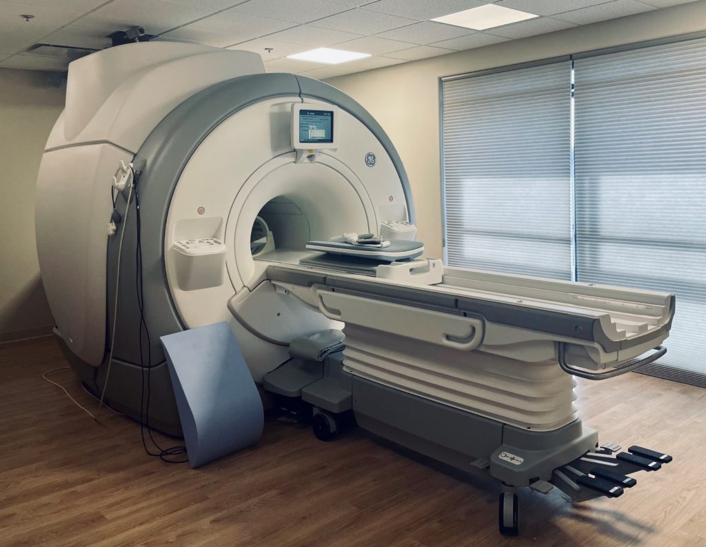
MRI: Unveiling Anatomy with Magnetic Fields
MRI employs powerful magnetic fields and radio waves to generate detailed images of the body’s soft tissues, including muscles, organs, and blood vessels. Unlike CT scans, which use ionizing radiation, MRI is considered a safe and non-invasive procedure.
MRI offers several advantages for abdominal imaging:
Superior Soft Tissue Imaging: MRI excels at visualizing soft tissues, providing detailed images of organs like the liver, pancreas, and kidneys.
No Radiation Exposure: Unlike CT scans, MRI does not involve ionizing radiation, making it a safer option for repeated imaging or for pregnant women.
Multiplanar Imaging: MRI can produce images in various planes, allowing for a comprehensive assessment of abdominal structures.
CT Scan: Capturing Cross-Sectional Views with X-Rays
CT scans utilize a series of X-rays to create detailed cross-sectional images of the body. These images are stacked together to form a 3D representation of the internal structures.

CT scans offer several advantages for abdominal imaging:
Fast Scanning Speed: CT scans are relatively quick, typically taking a few minutes to complete.
Detailed Bone Imaging: CT scans provide excellent images of bones, making them useful for detecting fractures or bone abnormalities.
Emergency Imaging: CT scans are often used in emergency situations due to their speed and ability to detect internal injuries.
MRI vs. CT Scan: Key Differences
The table below summarizes the key differences between MRI and CT scans of the abdomen:
| Feature | MRI | CT Scan |
|---|---|---|
| Imaging Principle | Magnetic fields and radio waves | Ionizing radiation |
| Strengths | Soft tissue imaging, no radiation exposure, multiplanar imaging | Fast scanning speed, detailed bone imaging, emergency imaging |
| Weaknesses | Long scanning time, claustrophobic feeling, not suitable for patients with metal implants | Radiation exposure, less detailed soft tissue imaging |
| Applications | Detecting soft tissue abnormalities, diagnosing liver and kidney diseases, evaluating tumors | Detecting internal injuries, assessing bone fractures, examining vascular disorders |
Choosing the Right Imaging Technique
The choice between MRI and CT scan for abdominal imaging depends on the specific clinical indications and patient considerations. Your doctor will carefully evaluate your symptoms, medical history, and any underlying conditions to determine the most appropriate imaging technique for your needs.
In general, MRI is preferred for visualizing soft tissues and diagnosing conditions that affect soft tissue structures, such as tumors, inflammation, or congenital anomalies. CT scans are often preferred for urgent situations, imaging of bones, and assessing potential internal injuries.
Conclusion
MRI and CT scans are both powerful diagnostic tools that play a crucial role in abdominal imaging. Understanding their unique strengths and limitations is essential for making informed decisions about the most suitable imaging technique for each patient. Consult with your doctor to determine the best course of action for your specific medical needs.